Screening for Glaucoma and Macular Degeneration in Scotts Valley: What Our Advanced Diagnostic Imaging Actually Sees

Screening for Glaucoma and Macular Degeneration in Scotts Valley: What Our Advanced Diagnostic Imaging Actually Sees

Screening for Glaucoma and Macular Degeneration in Scotts Valley: What Our Advanced Diagnostic Imaging Actually Sees

Screening for Glaucoma and Macular Degeneration in Scotts Valley: What Our Advanced Diagnostic Imaging Actually Sees

Screening for Glaucoma and Macular Degeneration in Scotts Valley: What Our Advanced Diagnostic Imaging Actually Sees

Screening for Glaucoma and Macular Degeneration in Scotts Valley: What Our Advanced Diagnostic Imaging Actually Sees

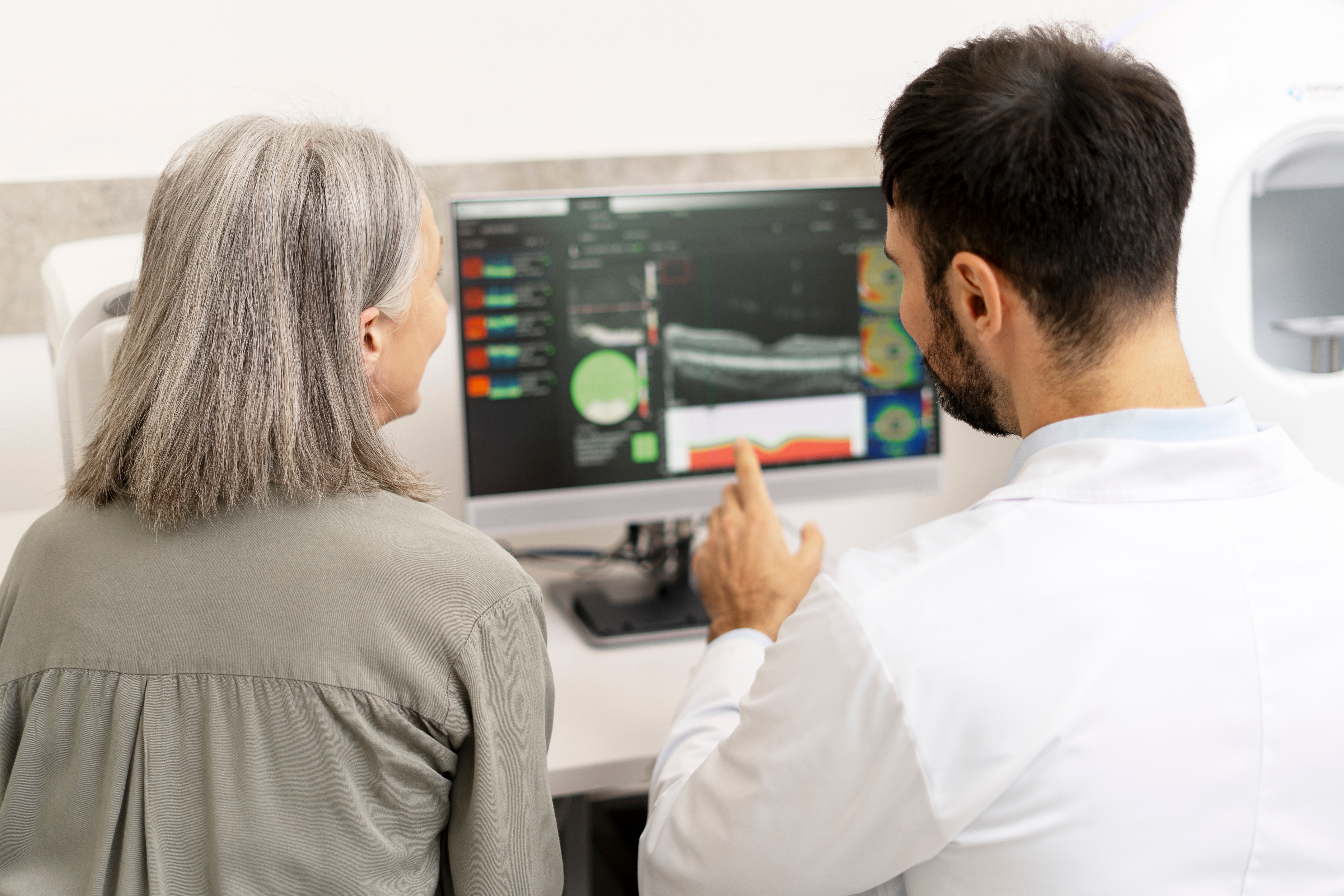
Glaucoma and macular degeneration can develop quietly, sometimes before noticeable vision changes appear. That is why a comprehensive eye exam involves more than checking whether you can read the smallest line on an eye chart.
At Giannotti Vision Care in Scotts Valley, we use advanced diagnostic technology to examine the retina, macula, optic nerve, and other structures inside the eye. Imaging can reveal subtle changes that may require closer monitoring, helping us build a more complete picture of your eye health.
What Does OCT Imaging Actually Show?
Optical Coherence Tomography, or OCT, produces high-resolution cross-sectional images of the retina and optic nerve. Think of it as allowing us to examine individual layers of tissue rather than only looking at the surface.
For glaucoma screening, OCT can measure structures around the optic nerve and help identify thinning that may be associated with glaucoma. For macular degeneration, it can provide detailed images of the macula and reveal structural changes within the retinal layers.
Because the images can be compared over time, OCT is also useful for monitoring whether suspicious findings remain stable or continue changing.
How Does Optomap Retinal Imaging Help?
Giannotti Vision Care also uses the Optomap Retinal Exam, which captures an ultra-widefield image of the retina.
This gives us a broad view of the back of the eye and creates a digital image that can become part of your health record. Depending on the individual patient, retinal imaging may help us document and monitor:
The optic nerve and surrounding retinal tissue
The macula responsible for detailed central vision
Retinal blood vessels
Changes associated with certain retinal diseases
Peripheral retinal abnormalities
Differences that develop between future eye exams
Optomap imaging can provide valuable information, but your optometrist will determine what combination of imaging and examination techniques is appropriate for your eyes.
What Are We Looking for With Glaucoma?
Glaucoma damages the optic nerve and often progresses without obvious early symptoms. By the time someone notices peripheral vision loss, permanent damage may already have occurred.
That is why glaucoma evaluation involves several pieces of information rather than one test alone. Along with OCT imaging, we may measure eye pressure, evaluate the appearance of the optic nerve, measure corneal thickness with pachymetry, and perform computerized visual field testing.
Visual field testing is especially important because it measures how well you can see across different areas of your field of vision. OCT shows us structural information, while the visual field test helps us understand how your vision is functioning.
What Does Imaging Reveal About Macular Degeneration?
Age-related macular degeneration affects the macula, the small central portion of the retina responsible for detailed vision used for reading, recognizing faces, and driving.
Early AMD may not immediately produce obvious symptoms. Advanced retinal imaging can help us examine the macula for structural changes and document findings so we can monitor them over time.
If you notice straight lines appearing wavy, reduced central clarity, dark areas in your central vision, or difficulty recognizing faces, schedule an eye examination promptly rather than waiting for your next routine visit.
Do You Need Every Imaging Test at Every Exam?
Not necessarily. Your age, family history, medical conditions, eye pressure, symptoms, previous findings, and other risk factors help determine which diagnostic tests are appropriate.
Advanced imaging is most useful when it supports a comprehensive evaluation rather than replacing one. By combining examination findings with detailed retinal and optic nerve imaging, we can determine whether your eyes appear healthy or whether additional monitoring is needed.
If it is time to take a closer look at your eye health, schedule a comprehensive eye exam with Giannotti Vision Care at 221 Mt Hermon Rd, Suite G, Scotts Valley, CA 95066 by calling (831) 438-4482. Our advanced diagnostic imaging can help us screen for glaucoma, macular degeneration, and other eye health changes before they have a greater impact on your vision.


Our Services
Contact Info
Hours of Operation
- Monday 8:30 AM - 12:00 PM
1:00 PM - 5:00 PM - Tuesday 8:30 AM - 12:00 PM
1:00:PM - 5:00PM - Wednesday 8:30 AM - 12:00 PM
1:00:PM - 5:00PM - Thursday 8:30 AM - 12:00 PM
1:00:PM - 5:00PM - Friday 8:30 AM - 12:00 PM
- Saturday Closed
- Sunday Closed
© 2026 Giannotti Vision Care, Optometry. All rights Reserved. Accessibility Statement - Privacy Policy - Sitemap
Powered by:
